Purbanchal Cancer Hospital has introduced advanced PET/CT (Positron Emission Tomography / Computed Tomography) imaging, a rapidly growing diagnostic modality that has made a tremendous impact on the care of patients with cancer and other medical conditions.
PET scans use a small amount of a radioactive substance, known as a tracer, to detect differences between healthy tissue and diseased tissue based on metabolic activity.
The most commonly used tracer is 18F-FDG (Fluoro-deoxy-glucose), which accumulates in cells with increased metabolic activity.
The combination of PET and CT provides complete information about both:
➤ Structure (CT) – detailed anatomical images
➤ Function (PET) – metabolic and cellular activity
This fusion allows precise localization and characterization of disease in the human body.
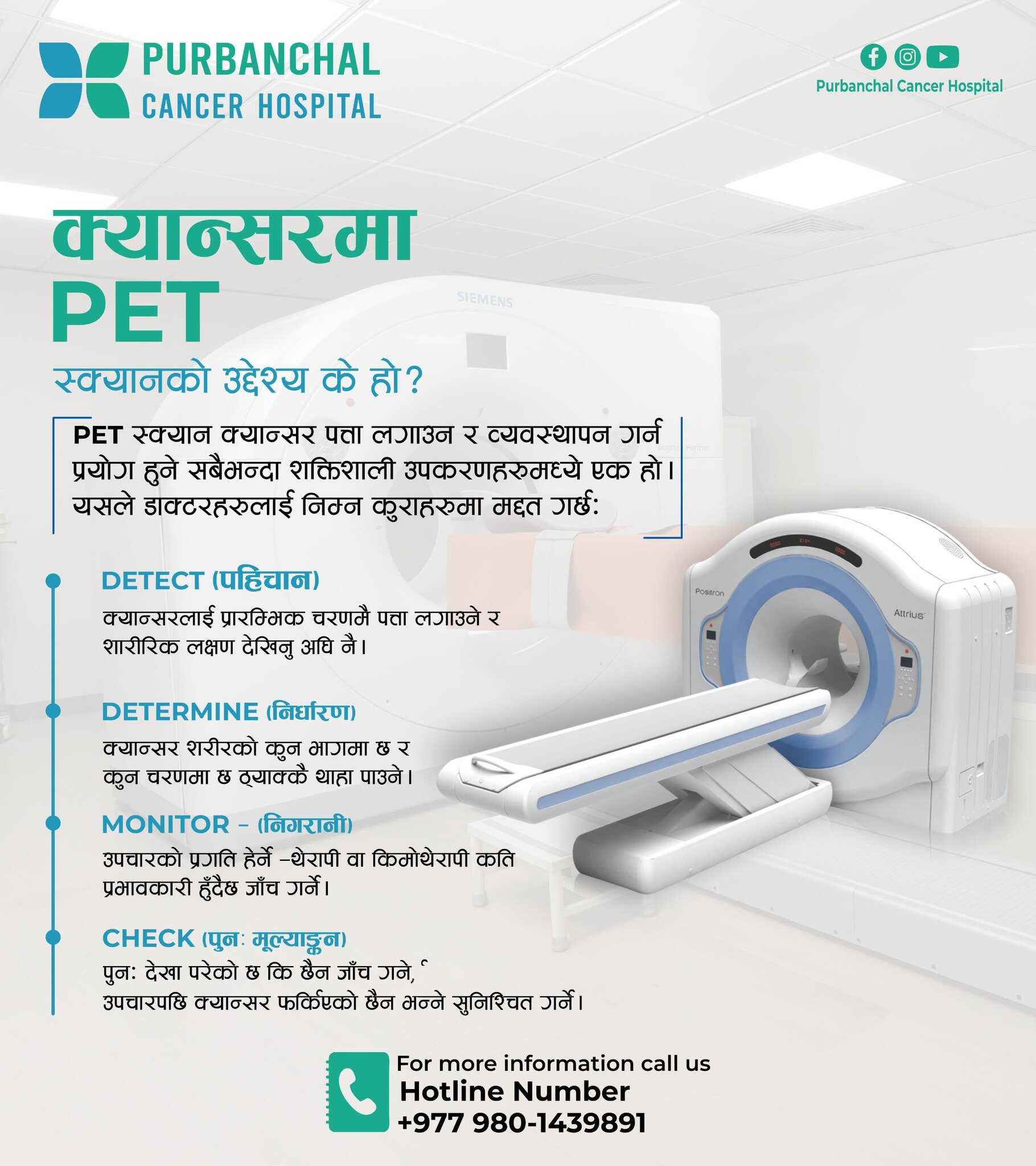
1. Oncological Applications (>95%)
PET/CT is widely used in the evaluation and management of various cancers, including:
➤ Lymphoma
➤ Head and Neck Cancers
➤ Lung Cancer
➤ Breast Cancer
➤ Renal Cell Carcinoma (RCC)
➤ Gynaecological Malignancies
➤ Metastatic Cancers
➤ Carcinoma of Unknown Primary
Useful for:
➤ Diagnosis of malignancy
➤ Staging of various cancers
➤ Response evaluation after chemotherapy, radiotherapy, surgery, or immunotherapy
2. Non-Oncological Applications (<5%)
Neurological Applications
PET/CT is used in the evaluation of epilepsy, dementia (including Alzheimer’s disease), and other neurodegenerative disorders by assessing brain metabolism and function.
Cardiac Applications
PET/CT helps in assessing myocardial viability and evaluating cardiac inflammation or infections, aiding in accurate cardiac diagnosis and treatment planning.
Infection and Inflammatory Conditions
PET/CT is valuable in detecting occult infections, evaluating fever of unknown origin, and assessing inflammatory diseases by identifying areas of increased metabolic activity.